
[:en]
Plasma Mole Removal Case Study
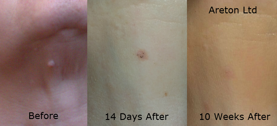
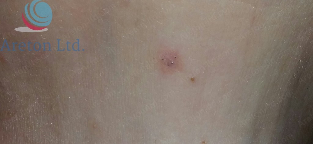
As seen in middle of the picture, 14 days after the procedure, the area is slightly red and it has a different texture. The treated area blended well 10 weeks after the treatment.
The following video will show you the procedure, before, the healing up until 10 weeks after the procedure. The video will provide you with an overview of the entire case study and will emphasize the important points you can learn from this experience.
The subject was approximately 45 years of age at the time of the treatment.
The mole was benign and removed for aesthetic reasons only, it was neither inflamed nor red.
The objective of the aesthetic practitioner was to level off the mole with the surrounding skin area in order to achieve the best possible results.
The numbing product used, was topical anaesthetic (10% lidocaine) applied with occlusion for one hour. Remember, the active ingredients contained in the topical numbing porducts available "over the counter" in Europe, can fary vary. In the specific country 10% lidocaine formulation was available "over the counter". In most European countries the formulation available without a prescription range between 2.5% and 5% lidocaine.
Although the particular numbing product was applied for a relative long time (one hour) to achieve the desired numbing effect, this does not mean that the only fast numbing alternative is injectable local anaesthetic. In fact, in most cases, there is no need to use injectable local anaesthetics for this types of procedures in order to achieve a fast numbing action of the area. In Europe, medical practitioners can use strong topical anaesthetic formulations which achieve the complete numbing of the area in 5 to 10 minutes, sometimes even without the use of occlusion.
As seen the skin is slightly pinker soon after the scabs has fallen off (during the long-term healing). This is a normal reaction after mole removal of any other minor skin wounds. The area will eventually blend with the surrounding skin, leading to seamless results in most cases. Always bear in mind that the area doe not always blend perfectly with the surrounding skin. Sometimes a minor permanent scars may result, despite the way the treatment was carried out.
In this case like in most others, 10 weeks after the treatment the result was almost seemless meaning that it was difficult to tell where the mole was previously located.
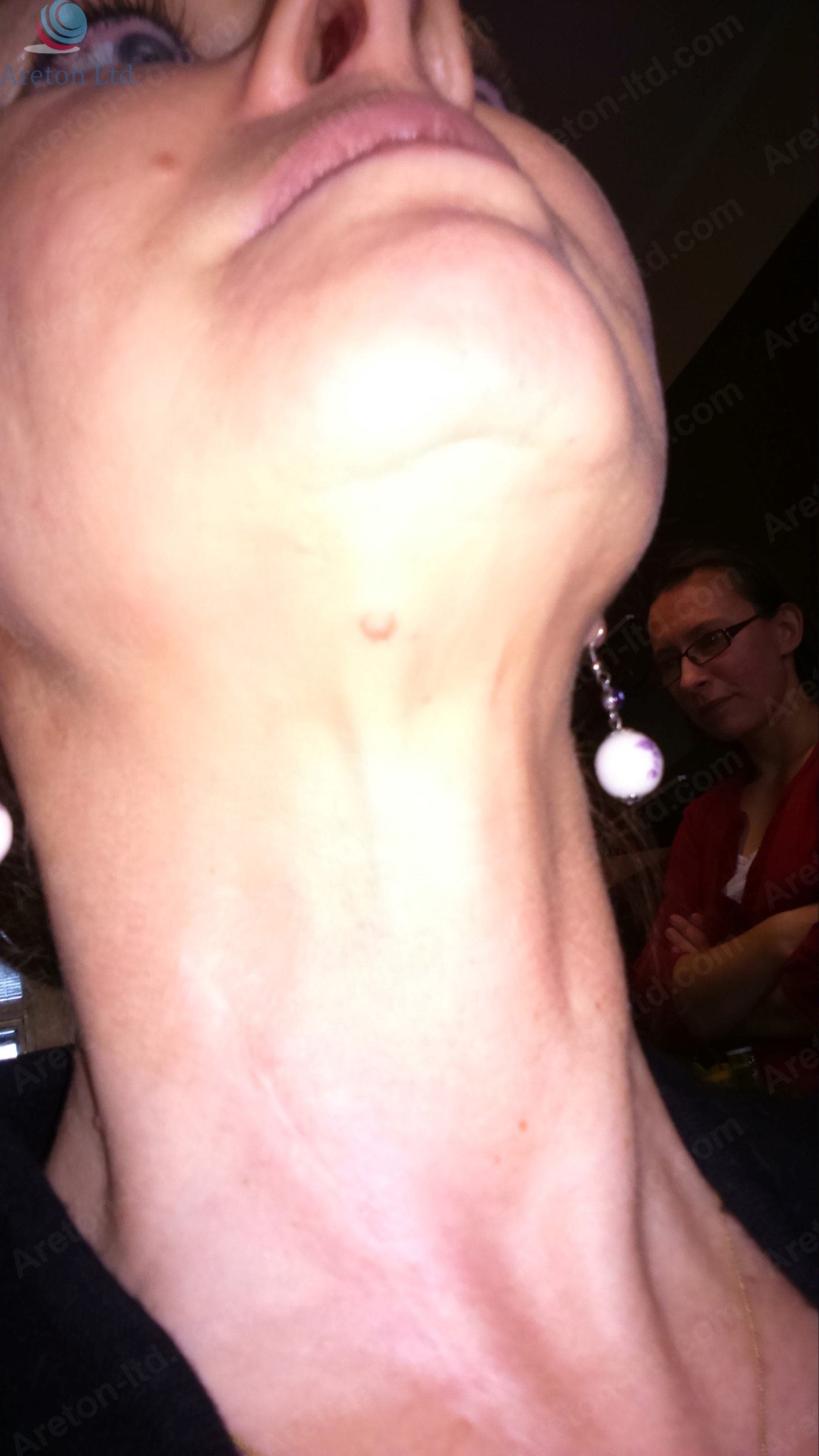
This picture shows where the mole was previously located.
Below you can appreciate a number of before pictures taken right before performing the plasma removal procedure. The mole was not subject to any change over time, this also confirmed that the lesion was benign.
[gallery_bank type="images" format="thumbnail" title="true" desc="true" responsive="true" display="all" sort_by="sort_order" animation_effect="bounce" album_title="true" album_id="11"]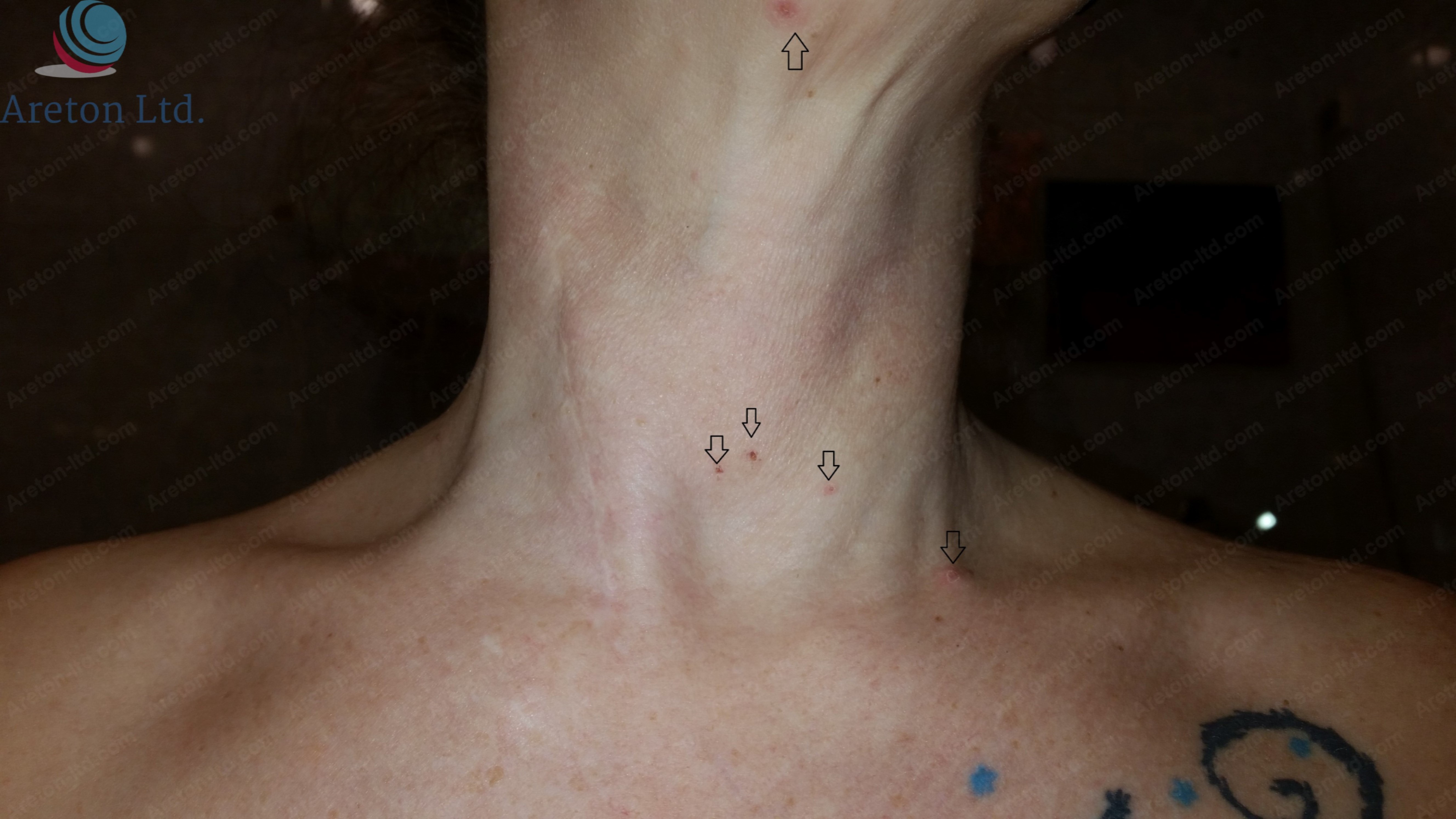
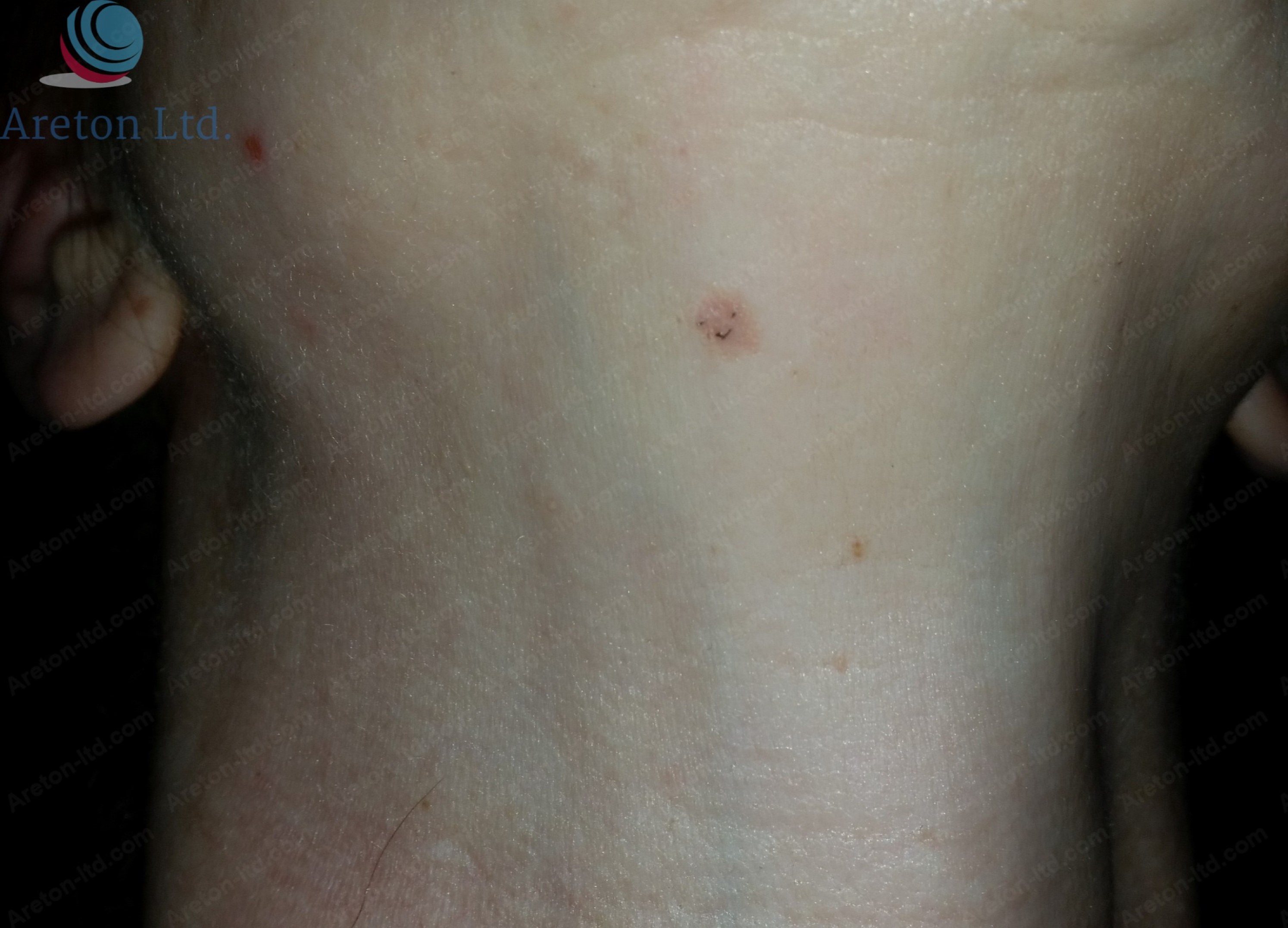
This picture shows the healing still in progress a few days after the plasma mole removal treatment. The arrows indicate where the plasma treatment was carried out. The mole in question in this case study is in the uppermost part of the picture and it is hardly visible due to the way this particular picture was taken at the time. However this picture shows how the treated area looks before the scabbing takes place.

Picture without the arrow indicators.
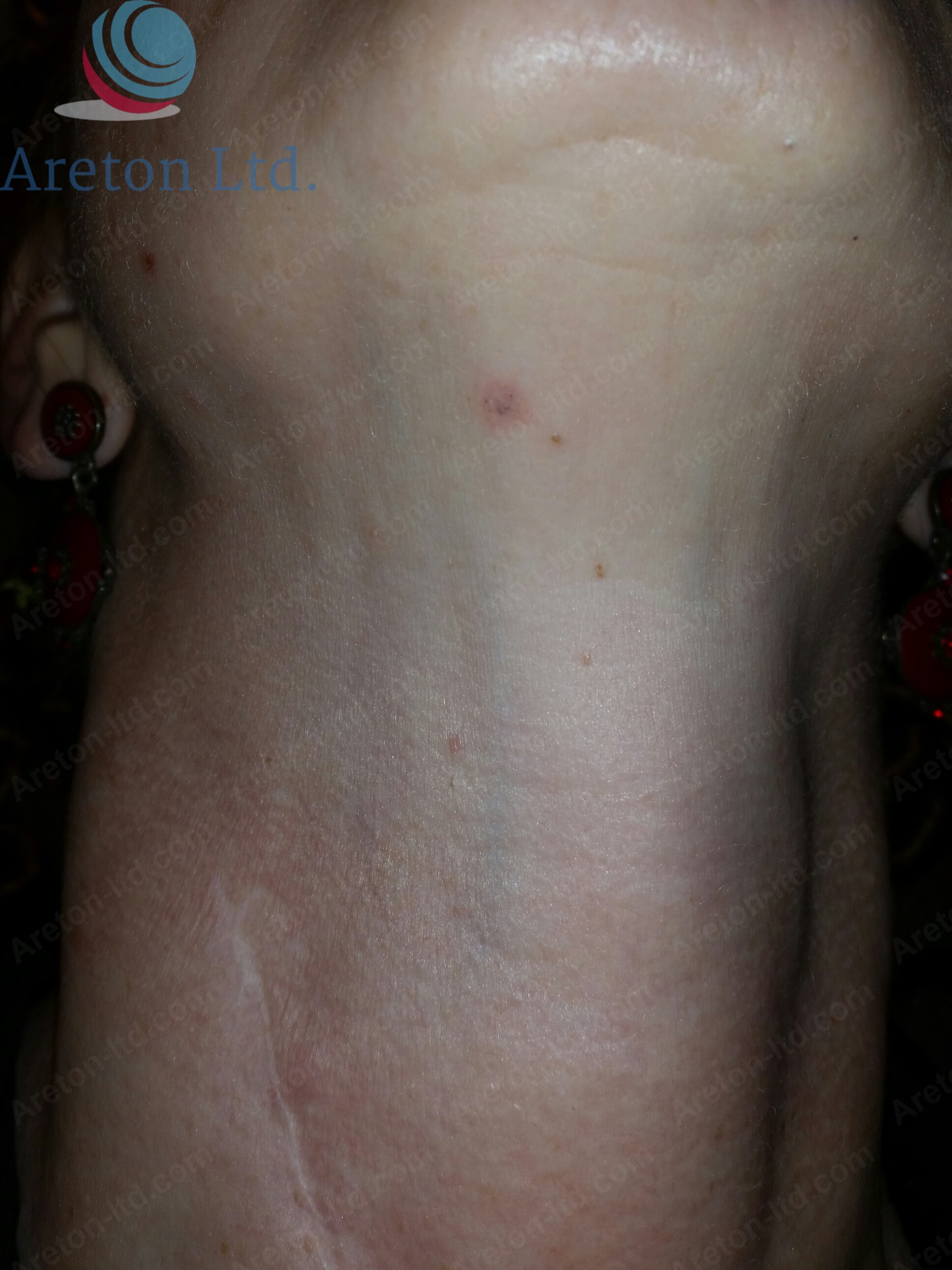
Area 10 Days After the Procedure.
The new skin is pinker and has a different texture to the surrounding skin. The area will eventually blend with the surrounding skin leading to seamless results.
14 Days after the procedure no major change occurred as the area is now subject to long-term healing. The area will change over time but the process will be rather slow.
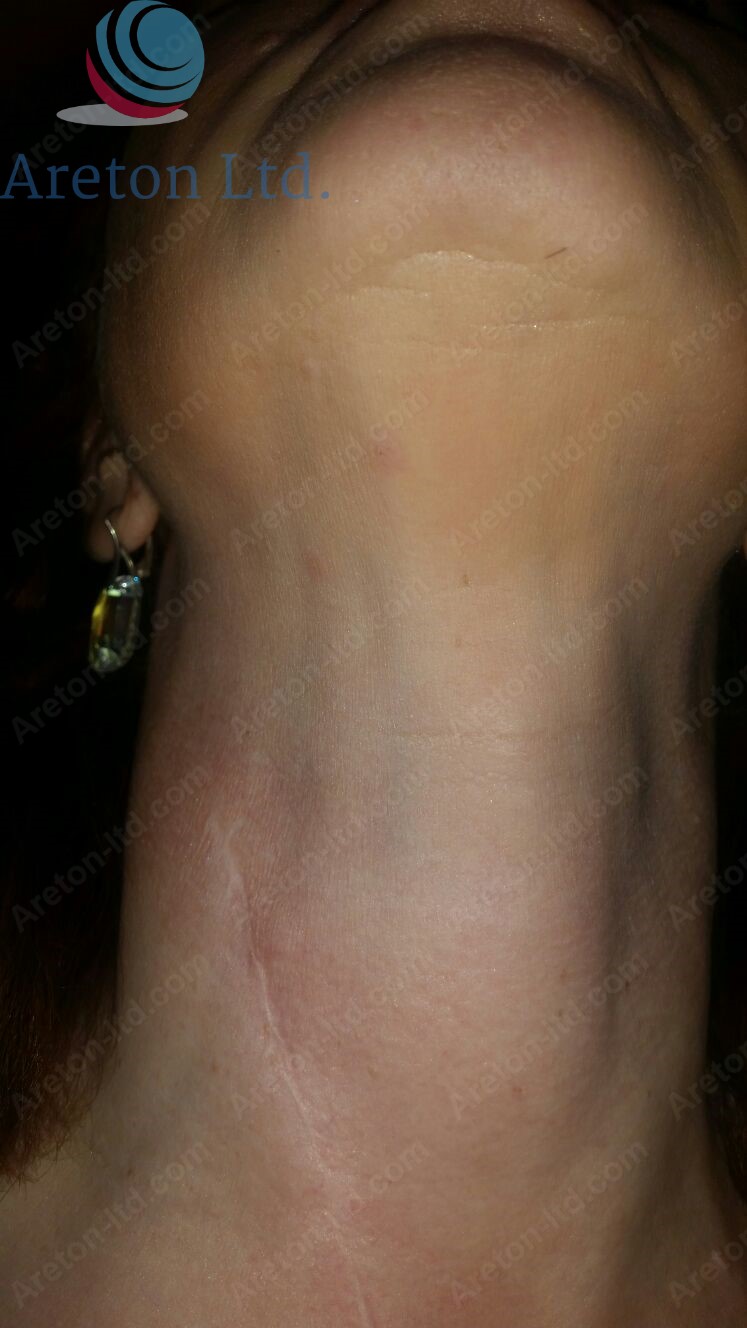
Area 10 weeks after the procedure. The location of the previous mole is hardly visible. In this case it is very likely the results will be complete seamless after a few more weeks.
In the following gallery you will see the most important pictures of this case study.
[gallery_bank type="images" format="thumbnail" title="true" desc="true" responsive="true" display="all" sort_by="sort_order" animation_effect="bounce" album_title="true" album_id="12"]
[:es]
Estudio de remoción de lunares con plasma
Como se ve en la mitad de la imagen, 14 días después del procedimiento, el área es ligeramente roja y tiene una textura diferente. El área tratada se mezcló bien 10 semanas después del tratamiento.
El siguiente video le mostrará el procedimiento, antes, la recuperación hasta 10 semanas después del procedimiento. El video le proporcionará una visión general de todo el estudio y enfatizará los puntos importantes que puede aprender de esta experiencia.
El sujeto tenía aproximadamente 45 años de edad en el momento del tratamiento.
El lunar era benigno y se eliminó solo por razones estéticas, no estaba inflamado ni rojo.
El objetivo del profesional de la estética era nivelar el lunar con el área de la piel circundante para lograr los mejores resultados posibles.
El producto anestésico utilizado fue anestesia tópica (lidocaína al 10%) aplicada con oclusión durante una hora. Recuerde, los ingredientes activos contenidos en los productos de anestesia tópica disponibles "de venta libre" en Europa pueden variar. En el país específico, la formulación de lidocaína al 10% estaba disponible "de venta libre". En la mayoría de los países europeos, la formulación disponible sin receta varía entre 2,5% y 5% de lidocaína..
Aunque el producto de adormecimiento en particular se aplicó durante un tiempo relativamente largo (una hora) para lograr el efecto de anestesia deseado, esto no significa que la única alternativa de adormecer rápido sea el anestésico local inyectable. De hecho, en la mayoría de los casos, no hay necesidad de usar anestésicos locales inyectables para este tipo de procedimientos, para lograr una rápida acción de adormecimiento del área. En Europa, los médicos pueden usar fuertes formulaciones anestésicas tópicas que logran el adormecimiento completo del área en5 a 10 minutos, a veces incluso sin el uso de oclusión..
Como se ve, la piel se torna un poco más rosada poco después de que se hayan caído las costras (durante la cicatrización a largo plazo). Esta es una reacción normal después de la eliminación de lunares de cualquier otra herida cutánea menor. El área eventualmente se mezclará con la piel circundante, lo que llevará a resultados perfectos en la mayoría de los casos. Siempre tenga en cuenta que el área no siempre se mezcla perfectamente con la piel circundante. A veces, se pueden producir cicatrices permanentes menores, a pesar de la forma en que se realizó el tratamiento.
En este caso, como en la mayoría de los otros, 10 semanas después del tratamiento, el resultado fue casi aparente, lo que significa que era difícil saber dónde se encontraba el lunar anteriormente.
Esta imagen muestra dónde se localizó el lunar anteriormente..
A continuación, puede apreciar una serie de fotografías tomadas antes de realizar el procedimiento de extracción de plasma. El lunar no estuvo sujeto a ningún cambio en el tiempo, esto también confirmó que la lesión era benigna.
[gallery_bank type="images" format="thumbnail" title="true" desc="true" responsive="true" display="all" sort_by="sort_order" animation_effect="bounce" album_title="true" album_id="11"]Esta imagen muestra la curación aún en progreso unos días después del tratamiento de remoción de lunares con plasma. Las flechas indican dónde se realizó el tratamiento con plasma. El lunar en cuestión en este estudio se encuentra en la parte superior de la imagen y apenas es visible debido a la forma en que se tomó esta imagen en particular en ese momento. Sin embargo, esta imagen muestra cómo se ve el área tratada antes de que tenga lugar la formación de costras.
Imagen sin los indicadores de flecha.
Área 10 días después del procedimiento.
La nueva piel es más rosada y tiene una textura diferente a la piel circundante. El área eventualmente se mezclará con la piel circundante dando lugar a resultados sin problemas.
14 días después del procedimiento, no se produjeron cambios importantes, ya que el área ahora está sujeta a una curación a largo plazo. El área cambiará con el tiempo, pero el proceso será bastante lento.
Área 10 semanas después del procedimiento. La ubicación del lunar anterior es apenas visible. En este caso, es muy probable que los resultados se completen sin problemas después de unas semanas más.
En la siguiente galería, verá las imágenes más importantes de este estudio puntual.
[gallery_bank type="images" format="thumbnail" title="true" desc="true" responsive="true" display="all" sort_by="sort_order" animation_effect="bounce" album_title="true" album_id="12"]
[:]